AtlasXomics Service
Work with our experts to map the epigenome in tissue
1. Meet with our experts to understand biological question and region of interest
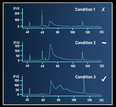
2. Sample optimization/ qualification to determine feasibility of sample or antibody target
3. Spatial analysis, sequencing and standard data report with raw data
4. Visualize processed data in our interactive shinyApp
Send your samples to AtlasXomics for tissue optimization and comprehensive spatial epigenome analysis. Our team will deliver a detailed report, including both raw and processed data, for your review and further insights.
Tissue Requirements
•OCT embedded fresh frozen samples, stored at -80C
•Mount 7-10 μm tissue sections onto 1”x3” slides coated for tissue adhesion
•Each tissue region of interest should be centered on the slide (please communicate to AtlasXomics team your area of interest)
•One region of interest per slide (can contain multiple sections if they are within the region of interest)
Region for chip placement on standard 1x3in slide


